
Sajarah aplikasi sareng peran mikroskop bedah dina bedah saraf
Dina sajarah bedah saraf, aplikasi tinamikroskop bedahmangrupikeun simbol anu inovatif, maju tina jaman bedah saraf tradisional anu ngalaksanakeun operasi handapeun panon taranjang ka jaman bedah saraf modéren anu ngalaksanakeun operasi dinamikroskopSaha sareng iraha ngalakukeunanamikroskop operasimimiti dianggo dina bedah saraf? Naon peran anu parantosmikroskop bedahmaénkeun peran dina kamekaran bedah saraf? Kalayan kamajuan élmu pangaweruh sareng téknologi, bakalMikroskop operasiNaha ieu téh kudu diganti ku alat-alat anu leuwih canggih? Ieu pertanyaan anu kudu diwaspadai ku unggal ahli bedah saraf sarta nerapkeun téknologi sarta instrumen pangénggalna kana widang bedah saraf, pikeun ningkatkeun kamampuh bedah bedah saraf.
1. Sajarah Aplikasi Mikroskop dina Widang Médis
Dina fisika, lénsa kacamata nyaéta lénsa konvéks kalayan struktur tunggal anu gaduh éfék pembesar, sareng pembesaranana diwatesan, katelah kacamata pembesar. Dina taun 1590, dua urang Walanda masang dua pelat lénsa konvéks di jero laras silinder anu ipis, sahingga nimukeun alat pembesar struktur komposit anu munggaran di dunya:mikroskopSaatos éta, struktur mikroskop terus ningkat, sareng pembesaranna terus ningkat. Dina waktos éta, para ilmuwan utamina nganggo ieumikroskop kompositpikeun niténan struktur leutik sasatoan sareng pepelakan, sapertos struktur sél. Saprak pertengahan dugi ka akhir abad ka-19, kacamata pembesar sareng mikroskop laun-laun diterapkeun dina widang kadokteran. Mimitina, ahli bedah nganggo kacamata pembesar gaya kacamata kalayan struktur lénsa tunggal anu tiasa disimpen dina jembatan irung pikeun operasi. Dina taun 1876, dokter Jerman Saemisch ngalaksanakeun operasi "mikroskopis" munggaran di dunya nganggo kaca pembesar kacamata majemuk (jenis operasi henteu dipikanyaho). Dina taun 1893, perusahaan Jerman Zeiss nimukeunmikroskop binokuler, utamina dianggo pikeun observasi ékspériméntal di laboratorium médis, ogé pikeun observasi lési kornea sareng bilik anterior dina widang oftalmologi. Dina taun 1921, dumasar kana panalungtikan laboratorium ngeunaan anatomi ceuli bagian jero sato, ahli otolaryngologi Swédia Nylen nganggo alat anu tetepmikroskop bedah monokulardirancang sareng diproduksi ku anjeunna nyalira pikeun ngalaksanakeun operasi otitis média kronis dina manusa, anu mangrupikeun bedah mikro anu sajati. Sataun ti harita, dokter Nylen anu unggul, Hlolmgren, ngenalkeun amikroskop bedah binokulardiproduksi ku Zeiss di kamar operasi.
Anu mimitiMikroskop operasingagaduhan seueur kakurangan, sapertos stabilitas mékanis anu goréng, henteu mampuh gerak, cahaya tina sumbu anu béda sareng pemanasan lénsa obyektif, widang pembesaran bedah anu sempit, jsb. Ieu sadayana alesan anu ngawatesan aplikasi anu langkung lega tinamikroskop bedahDina tilu puluh taun ka hareup, kusabab interaksi positif antara ahli bedah sarengpabrik mikroskop, kinerja tinamikroskop bedahterus ditingkatkeun, sarengmikroskop bedah binokular, mikroskop anu dipasang dina hateup, lénsa zum, iluminasi sumber cahaya koaksial, panangan artikulasi anu dikontrol tekanan éléktronik atanapi cai, kontrol pedal suku, sareng saterasna sacara berturut-turut dikembangkeun. Dina taun 1953, perusahaan Jerman Zeiss ngahasilkeun sarangkaian khususmikroskop bedah pikeun otologi, utamana cocog pikeun operasi dina lesi anu jero sapertos ceuli tengah sareng tulang temporal. Sedengkeun kinerjamikroskop bedahterus ningkat, pola pikir ahli bedah ogé terus robih. Salaku conto, dokter Jerman Zollner sareng Wullstein netepkeun yénmikroskop bedahkedah dianggo pikeun operasi ngabentuk mémbran timpani. Saprak taun 1950-an, dokter mata laun-laun ngarobih prakték ngan ukur nganggo mikroskop pikeun pamariksaan mata sareng ngenalkeunmikroskop otosurgicalkana bedah mata. Saprak harita,Mikroskop operasiparantos seueur dianggo dina widang otologi sareng oftalmologi.
2. Aplikasi mikroskop bedah dina bedah saraf
Kusabab kakhususan bedah saraf, aplikasi tinamikroskop bedah dina bedah sarafrada telat tibatan dina otologi sareng oftalmologi, sareng ahli bedah saraf aktip diajar téknologi énggal ieu. Dina waktos éta,panggunaan mikroskop bedahutamina di Éropa. Dokter mata Amérika Perrit mimiti ngenalkeunmikroskop bedahti Éropa ka Amérika Serikat dina taun 1946, neundeun pondasi pikeun ahli bedah saraf Amérika pikeun ngagunakeunMikroskop operasi.
Tina sudut pandang ngahormatan ajén kahirupan manusa, téknologi, alat, atanapi instrumen énggal anu dianggo pikeun awak manusa kedah ngalaman ékspérimén awal ka sato sareng pelatihan téknis pikeun operator. Dina taun 1955, ahli bedah saraf Amérika Malis ngalaksanakeun operasi otak dina sato nganggomikroskop bedah binokularKurze, saurang ahli bedah saraf di Universitas California Kidul di Amérika Serikat, nyéépkeun sataun diajar téknik bedah ngagunakeun mikroskop di laboratorium saatos niténan bedah ceuli dina mikroskop. Dina bulan Agustus 1957, anjeunna hasil ngalaksanakeun bedah neuroma akustik ka murangkalih umur 5 taun ngagunakeunmikroskop bedah ceuli, anu mangrupikeun bedah mikro munggaran di dunya. Teu lami saatos éta, Kurze suksés ngalaksanakeun anastomosis saraf sublingual saraf wajah dina murangkalih nganggo amikroskop bedah, sareng pamulihan anak éta saé pisan. Ieu mangrupikeun bedah mikro anu kadua di dunya. Saatos éta, Kurze nganggo treuk pikeun ngangkutMikroskop operasika sababaraha tempat pikeun bedah saraf mikro, sareng nyarankeun pisan panggunaanmikroskop bedahka ahli bedah saraf anu sanés. Saatos éta, Kurze ngalaksanakeun operasi kliping aneurisma serebral nganggo amikroskop bedah(hanjakalna, anjeunna teu medalkeun artikel naon waé). Kalayan dukungan ti pasien neuralgia trigeminal anu diubaran ku anjeunna, anjeunna ngadegkeun laboratorium bedah saraf dasar tangkorak mikro munggaran di dunya dina taun 1961. Urang kedah teras émut kontribusi Kurze kana bedah mikro sareng diajar tina kawani na pikeun nampi téknologi sareng ideu énggal. Nanging, dugi ka awal taun 1990-an, sababaraha ahli bedah saraf di Cina henteu nampiMikroskop bedah sarafpikeun operasi. Ieu sanés masalah sarengMikroskop bedah sarafsorangan, tapi masalah sareng pamahaman idéologis ahli bedah saraf.
Dina taun 1958, ahli bedah saraf Amérika Donaghy ngadegkeun laboratorium panalungtikan sareng pelatihan bedah mikro munggaran di dunya di Burlington, Vermont. Dina tahap awal, anjeunna ogé ngalaman kabingungan sareng kasusah kauangan ti atasanna. Dina akademisi, anjeunna salawasna ngabayangkeun motong pembuluh darah kortikal pikeun langsung ngaluarkeun trombi tina pasien anu ngagaduhan trombosis serebral. Janten anjeunna damel bareng sareng ahli bedah vaskular Jacobson dina panalungtikan sato sareng klinis. Dina waktos éta, dina kaayaan panon taranjang, ngan ukur pembuluh darah alit kalayan diaméter 7-8 milimeter atanapi langkung anu tiasa dijahit. Pikeun ngahontal anastomosis tungtung-ka-tungtung pembuluh darah anu langkung lemes, Jacobson mimitina nyobian nganggo kaca pembesar gaya kacamata. Teu lami saatos éta, anjeunna émut nganggomikroskop bedah otolaringologipikeun operasi nalika anjeunna janten dokter residen. Janten, kalayan bantosan Zeiss di Jerman, Jacobson ngarancang mikroskop bedah operator ganda (Diploskop) pikeun anastomosis vaskular, anu ngamungkinkeun dua ahli bedah pikeun ngalakukeun operasi sacara babarengan. Saatos ékspérimén sato anu éksténsif, Jacobson nerbitkeun artikel ngeunaan anastomosis mikrosurgikal anjing sareng arteri non-karotid (1960), kalayan tingkat patensi anastomosis vaskular 100%. Ieu mangrupikeun makalah médis anu inovatif anu aya hubunganana sareng bedah saraf mikrosurgikal sareng bedah vaskular. Jacobson ogé ngarancang seueur instrumen bedah mikro, sapertos gunting mikro, wadah jarum mikro, sareng gagang instrumen mikro. Dina taun 1960, Donaghy suksés ngalaksanakeun trombektomi insisi arteri serebral dina amikroskop bedahpikeun pasien anu ngagaduhan trombosis serebral. Rhoton ti Amérika Serikat mimiti nalungtik anatomi otak dina mikroskop dina taun 1967, ngarintis widang anyar anatomi mikrosurgis sareng ngadamel kontribusi anu signifikan pikeun pamekaran mikrosurgis. Kusabab kaunggulan tinamikroskop bedahsareng kamajuan instrumen bedah mikro, beuki seueur ahli bedah anu resep nganggomikroskop bedahpikeun operasi. Sareng nerbitkeun seueur artikel anu aya hubunganana ngeunaan prosedur bedah mikro.
3. Aplikasi mikroskop bedah dina bedah saraf di Cina
Salaku urang Tionghoa perantauan anu patriotik di Jepang, Profesor Du Ziwei nyumbangkeun sumbangan domestik anu munggaranmikroskop bedah sarafsareng anu aya hubungananaalat bedah mikroka Departemen Bedah Saraf Rumah Sakit Afiliasi Suzhou Medical College (ayeuna Departemen Bedah Saraf Rumah Sakit Afiliasi Pertama Universitas Suzhou) dina taun 1972. Saatos uih deui ka Cina, anjeunna mimiti ngalaksanakeun operasi mikro sapertos aneurisma intrakranial sareng meningioma. Saatos diajar ngeunaan kasadiaanmikroskop bedah sarafsareng instrumen bedah mikro, Profesor Zhao Yadu ti Departemen Bedah Saraf Rumah Sakit Yiwu Beijing nganjang ka Profesor Du Ziwei ti Suzhou Medical College pikeun niténan panggunaanmikroskop bedahProfesor Shi Yuquan ti Rumah Sakit Shanghai Huashan sacara pribadi nganjang ka departemen Profesor Du Ziwei pikeun niténan prosedur bedah mikro. Hasilna, gelombang bubuka, pembelajaran, sareng aplikasi tinaMikroskop bedah sarafdipicu di pusat-pusat bedah saraf utama di Cina, nandakeun awal bedah saraf mikro Cina.
4. Pangaruh Bedah Mikro
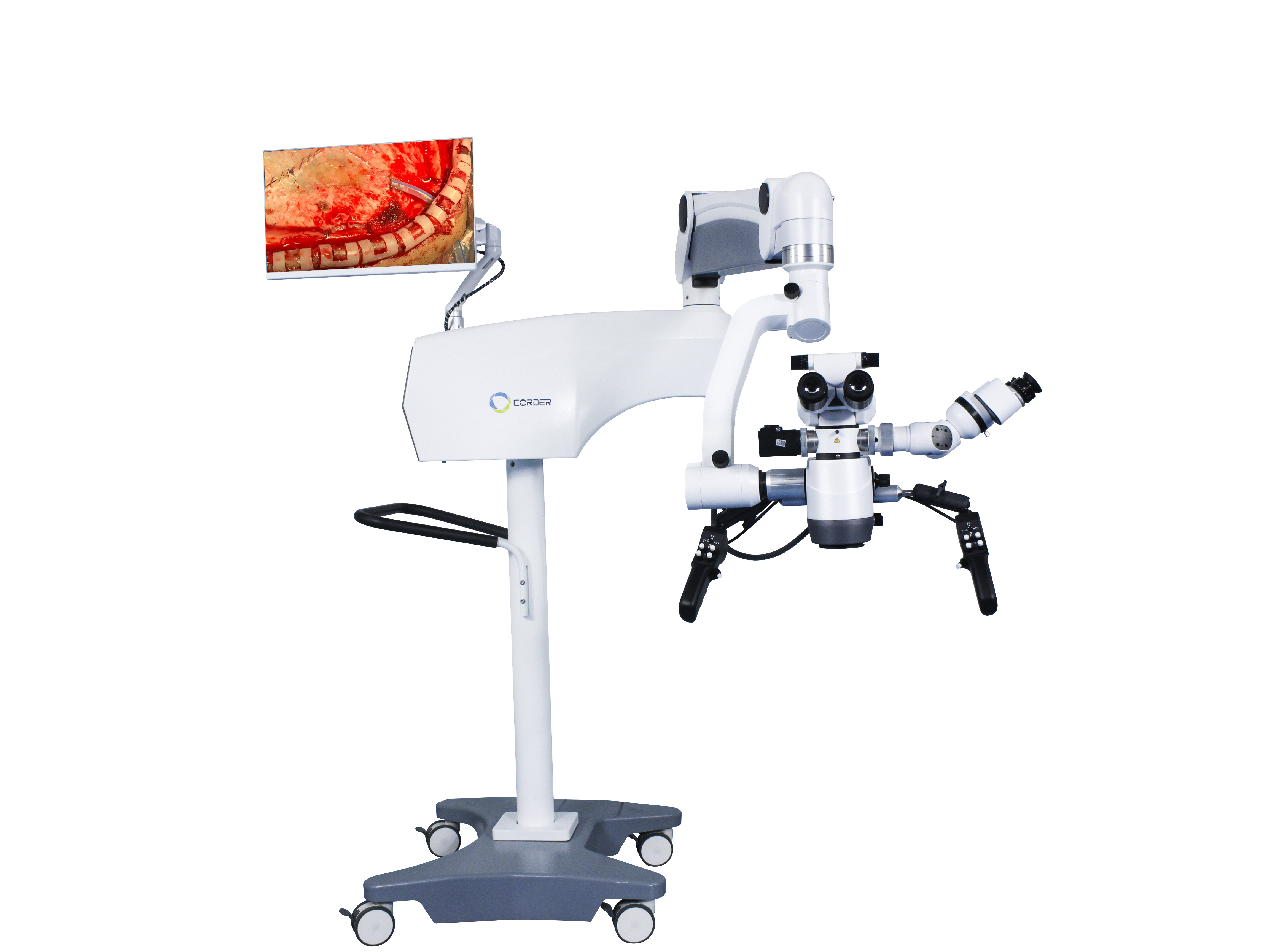
Kusabab panggunaanmikroskop bedah saraf, operasi anu teu tiasa dilakukeun ku panon taranjang janten tiasa dilakukeun dina kaayaan pembesaran 6-10 kali. Salaku conto, ngalaksanakeun operasi tumor hipofisis ngalangkungan sinus etmoidal tiasa ngaidentipikasi sareng miceun tumor hipofisis kalayan aman bari ngajaga kelenjar hipofisis normal; Operasi anu teu tiasa dilakukeun ku panon taranjang tiasa janten operasi anu langkung saé, sapertos tumor batang otak sareng tumor intrameduler tulang tonggong. Akademisi Wang Zhongcheng ngagaduhan tingkat mortalitas 10,7% pikeun operasi aneurisma serebral sateuacan nganggo amikroskop bedah sarafSaatos nganggo mikroskop dina taun 1978, tingkat mortalitas turun janten 3,2%. Tingkat mortalitas operasi malformasi arteriovenosa serebral tanpa nganggomikroskop bedahnyaéta 6,2%, sareng saatos 1984, kalayan panggunaan amikroskop bedah saraf, tingkat mortalitas turun jadi 1,6%. Panggunaanmikroskop bedah sarafngamungkinkeun tumor hipofisis diubaran ngaliwatan pendekatan transnasal transsphenoidal minimal invasif tanpa kedah kraniotomi, ngirangan tingkat mortalitas bedah tina 4,7% janten 0,9%. Pencapaian hasil ieu mustahil dina bedah panon kotor tradisional, jantenmikroskop bedahmangrupikeun simbol bedah saraf modéren sareng parantos janten salah sahiji alat bedah anu teu tiasa digentoskeun sareng teu tiasa digentos dina bedah saraf modéren.
Waktos posting: 09-Des-2024


