
Aplikasi mikroskop bedah gigi dina pengobatan panyakit pulpa sareng periapikal
Mikroskop bedahmibanda kaunggulan ganda nyaéta pembesaran sareng iluminasi, sareng parantos diterapkeun dina widang médis salami langkung ti satengah abad, ngahontal hasil anu tangtu.Mikroskop operasiloba dipaké sarta dimekarkeun dina bedah ceuli dina taun 1940 sarta dina bedah oftalmik dina taun 1960.
Dina widang kedokteran gigi,mikroskop bedahparantos diterapkeun kana tambalan huntu sareng perawatan restorasi ti mimiti taun 1960-an di Éropa. Aplikasi tinamikroskop operasidina endodontik sabenerna dimimitian dina taun 1990-an, nalika sarjana Italia Pecora mimiti ngalaporkeun panggunaanmikroskop bedah gigidina bedah endodontik.
Dokter gigi ngalengkepan pangobatan panyakit pulpa sareng periapikal dina amikroskop operasi gigiMikroskop bedah huntu tiasa ngagedekeun daérah lokal, niténan struktur anu langkung lemes, sareng nyayogikeun sumber cahaya anu cekap, ngamungkinkeun dokter gigi ningali struktur saluran akar sareng jaringan periapikal kalayan jelas, sareng mastikeun posisi bedah. Mikroskop ieu henteu ngan ukur ngandelkeun perasaan sareng pangalaman pikeun perawatan, sahingga ngirangan kateupastian perawatan sareng ningkatkeun kualitas perawatan pikeun panyakit pulpa sareng periapikal, ngamungkinkeun sababaraha huntu anu henteu tiasa dijaga ku metode tradisional pikeun nampi perawatan sareng pelestarian anu komprehensif.
A mikroskop huntudiwangun ku sistem iluminasi, sistem pembesaran, sistem pencitraan, sareng asesorisna. Sistem pembesaran diwangun ku eyepiece, tabung, lénsa objektif, pangatur pembesaran, jsb., anu sacara koléktif nyaluyukeun pembesaran.
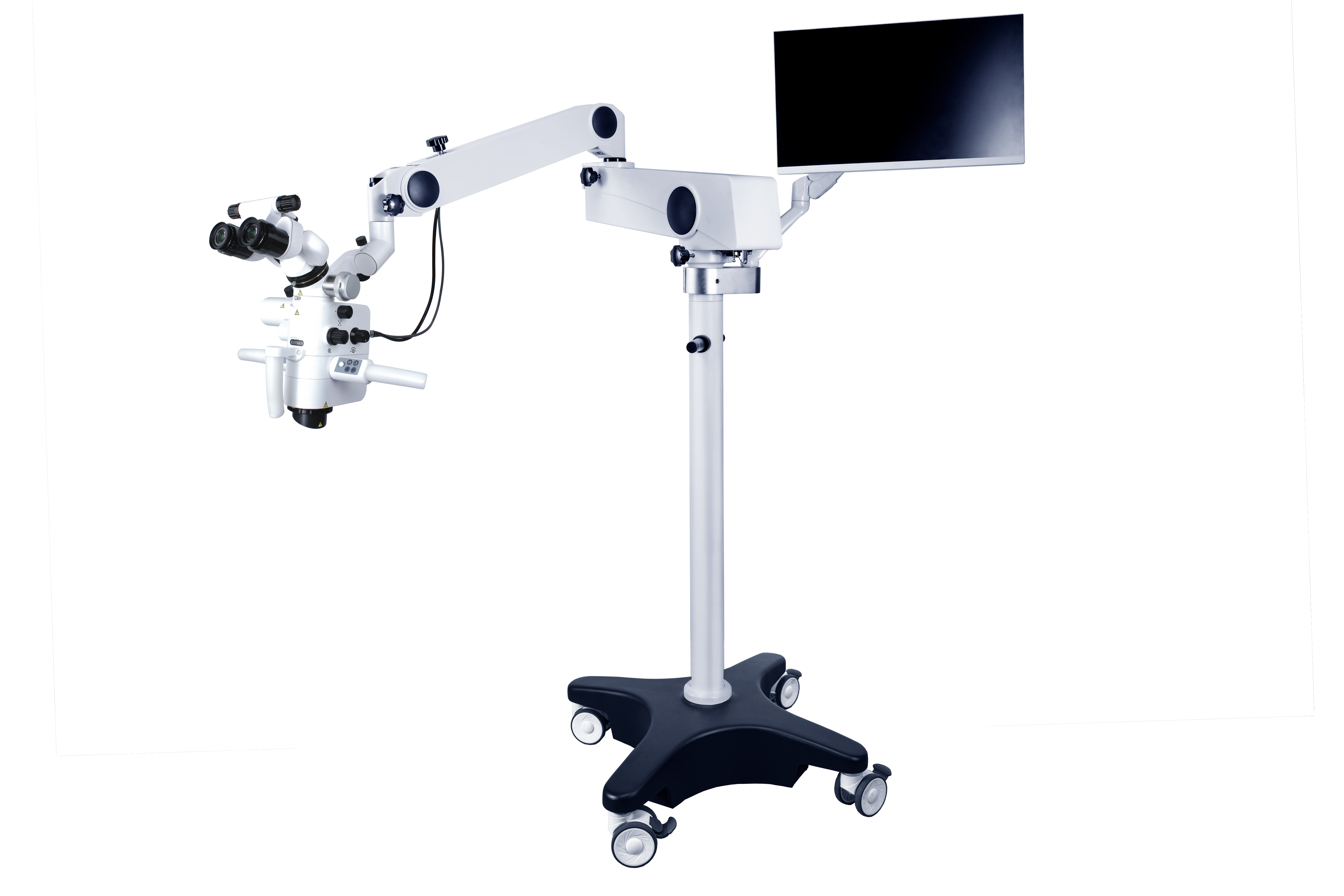
Nyandak CORDERMikroskop bedah gigi ASOM-520-DSalaku conto, pembesaran okuler aya dina rentang ti 10 × dugi ka 15 ×, kalayan pembesaran anu umum dianggo nyaéta 12,5X, sareng panjang fokus lénsa objektif aya dina kisaran 200 ~ 500 mm. Pangganti pembesaran gaduh dua modeu operasi: pangaturan tanpa léngkah listrik sareng pangaturan pembesaran kontinyu manual.
Sistem penerangan anumikroskop bedahdisayogikeun ku sumber cahaya serat optik, anu nyayogikeun katerangan paralel anu caang pikeun widang pandang sareng henteu ngahasilkeun kalangkang di daérah widang bedah. Nganggo lénsa binokular, dua panon tiasa dianggo pikeun observasi, ngirangan kacapean; Kéngingkeun gambar objék tilu diménsi. Salah sahiji metode pikeun ngarengsekeun masalah asistén nyaéta ku cara ngalengkepan eunteung asistén, anu tiasa nyayogikeun pandangan anu sami sareng ahli bedah, tapi biaya pikeun ngalengkepan eunteung asistén relatif luhur. Metode anu sanés nyaéta masang sistem kaméra dina mikroskop, nyambungkeunana kana layar tampilan, sareng ngamungkinkeun asistén ningali dina layar. Sakabéh prosés bedah ogé tiasa difoto atanapi dirékam pikeun ngumpulkeun rékaman médis pikeun pangajaran atanapi panalungtikan ilmiah.
Salila pangobatan panyakit pulpa sareng periapikal,mikroskop bedah gigitiasa dianggo pikeun nalungtik liang saluran akar, ngabersihkeun saluran akar anu kalsifikasi, ngalereskeun perforasi témbok saluran akar, mariksa morfologi saluran akar sareng efektivitas beberesih, miceun instrumen anu rusak sareng tumpukan saluran akar anu rusak, sareng ngalaksanakeunbedah mikroprosedur pikeun panyakit periapikal.
Dibandingkeun sareng bedah tradisional, kaunggulan bedah mikro kalebet: posisi anu tepat tina puncak akar; Réseksi bedah tradisional tulang gaduh rentang anu langkung ageung, sering langkung ageung atanapi sami sareng 10mm, sedengkeun karusakan tulang bedah mikro gaduh rentang anu langkung alit, kirang ti atanapi sami sareng 5mm; Saatos nganggo mikroskop, morfologi permukaan akar huntu tiasa dititénan kalayan leres, sareng sudut lamping motong akar kirang ti 10°, sedengkeun sudut lamping motong akar tradisional langkung ageung (45°); Kamampuh pikeun niténan isthmus antara saluran akar di tungtung akar; Tiasa nyiapkeun sareng ngeusian ujung akar sacara akurat. Salaku tambahan, éta tiasa mendakan landmark anatomis normal tina situs patah akar sareng sistem saluran akar. Prosés bedah tiasa difoto atanapi dirékam pikeun ngumpulkeun data pikeun tujuan klinis, pangajaran, atanapi panalungtikan ilmiah. Éta tiasa dianggap yénmikroskop bedah gigiMibanda nilai aplikasi sareng prospek anu saé dina diagnosis, pangobatan, pangajaran, sareng panalungtikan klinis panyakit pulpa gigi.
Waktos posting: 19-Des-2024


